Animal Models
Mark findings visually on animal models
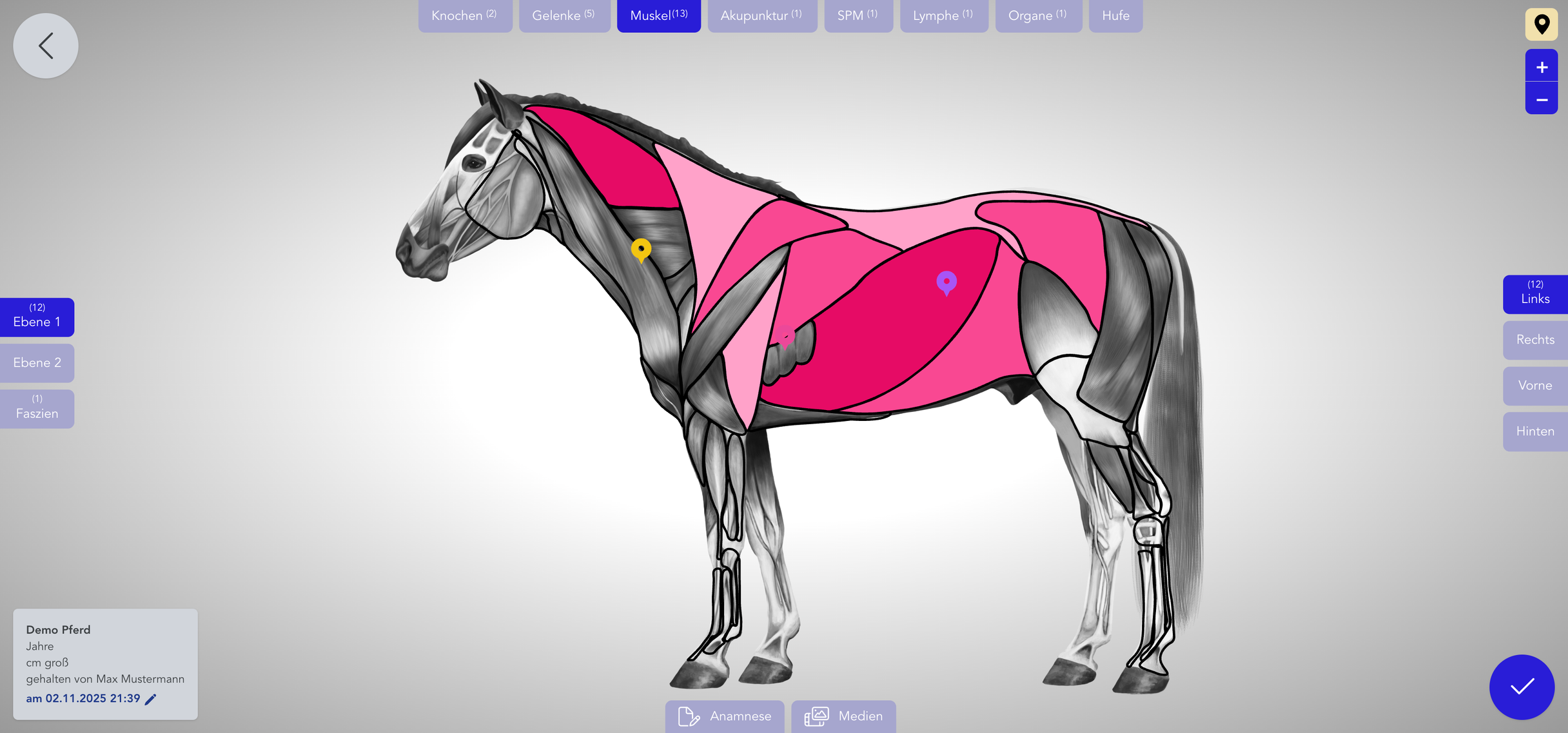
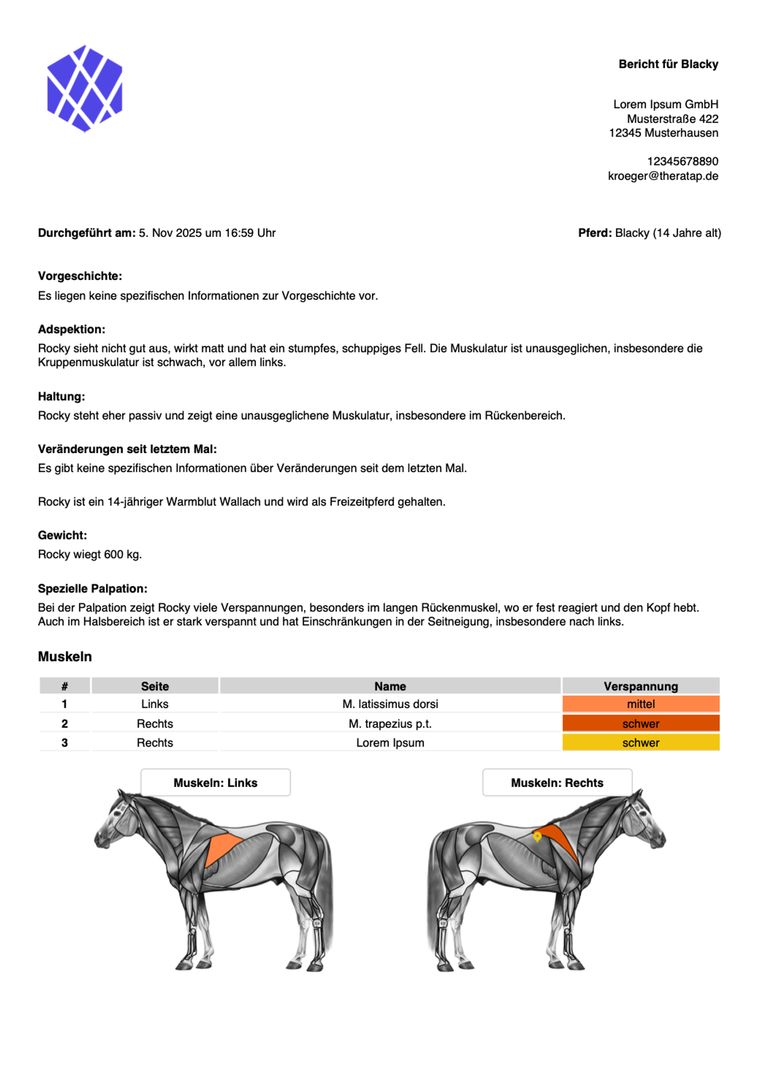
Mark findings directly on anatomical views of horse, dog, and cat. Colorful, precise, and digital. The markings are automatically saved and can be displayed in PDF documents.
Mark findings visually on animal models
Mark findings directly on anatomical views of horse, dog, and cat. Colorful, precise, and digital. The markings are automatically saved and can be displayed in PDF documents.
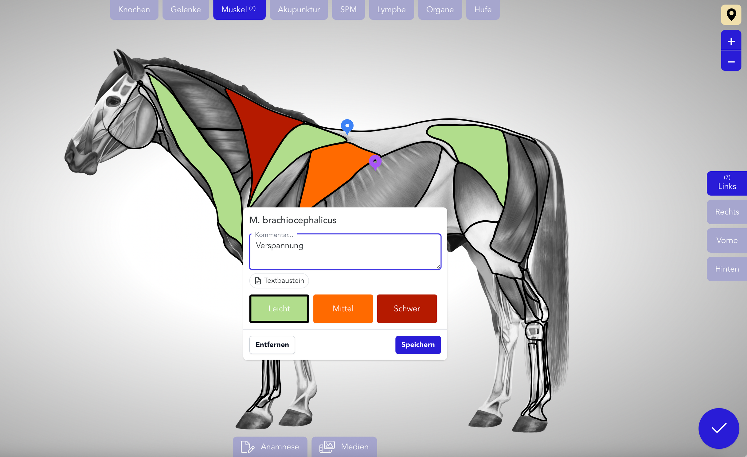
The animal models consist of multiple anatomical layers that enable even more precise findings. Each layer shows different structures – ideal for precise analyses and for comparing between different appointments.
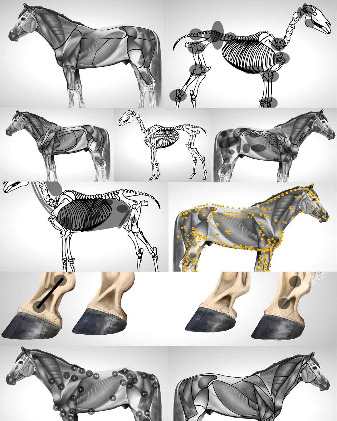
10 layers
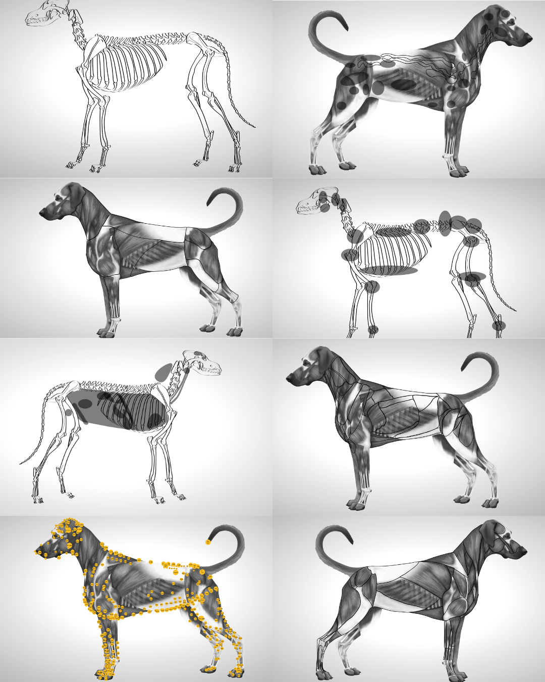
8 layers
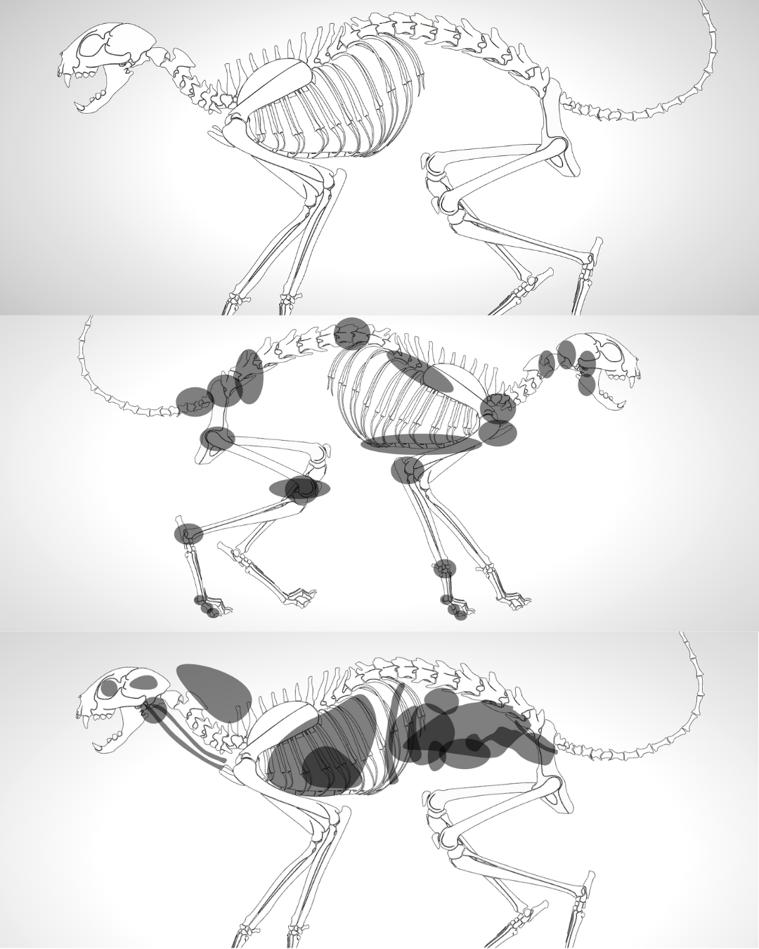
3 layers
You can switch between layers at any time to make exactly the structures visible that are relevant for your findings.
Visualize findings precisely
With TheraTap's animal models, you document findings visually and precisely. Use various tools, colors, and views for professional documentation.
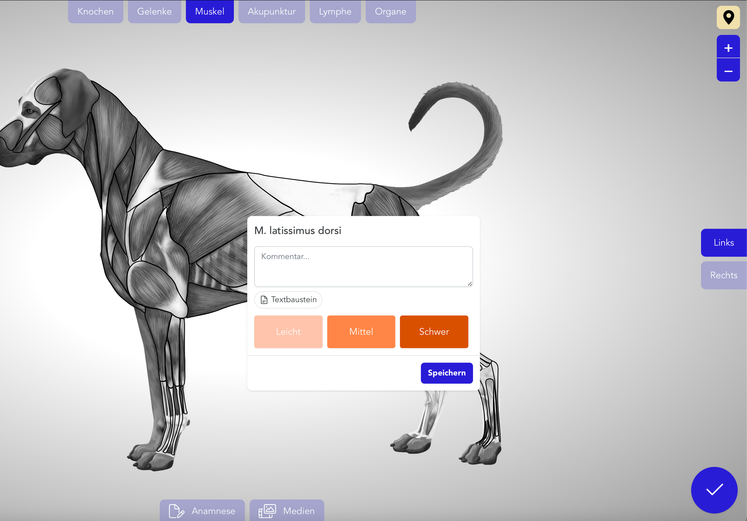
Mark exactly where a problem is located – without interpretation errors.
At the next appointment, you immediately see,
→ Where was the problem?
→ How has it developed?
Customers understand visual markers immediately.
Reports become clearer and misunderstandings disappear.
With TheraTap's animal models, you document findings faster and more understandably. Save time in documentation and focus on what really matters: your patients.
From Model to Documentation
With the animal models, you set up your visual findings in just a few clicks. Mark problem areas, add comments, and export everything directly to your findings report.
With TheraTap's animal models, you document findings faster and more understandably. Save time in documentation and focus on what really matters: your patients.
As soon as you start a treatment, TheraTap automatically loads the appropriate animal model based on the animal species. You can then mark directly on the anatomical model – with text labels, markers, and various colors. All markings are automatically saved.
Currently, detailed anatomical models are available for horse, dog, and cat. The appropriate model is automatically loaded based on the animal file.
Depending on the animal species, several anatomical views are available, e.g., left, right, front, and back. The markings per view are saved separately, so you can document each view individually.
Yes, you can define your own colors for different finding types in your settings. Each tool supports multiple colors that you can individually maintain.
Yes, all markings are automatically integrated into your findings report. The visual markings are displayed in the PDF – perfect for sharing with pet owners or veterinarians. In the MAX plan, the markings are even integrated as images.
The anatomical models themselves are predefined, but you can individually customize colors, tools, and labels. This way you create documentation that fits exactly to your workflow.