






Tap a muscle, enter the finding -- now in 3D
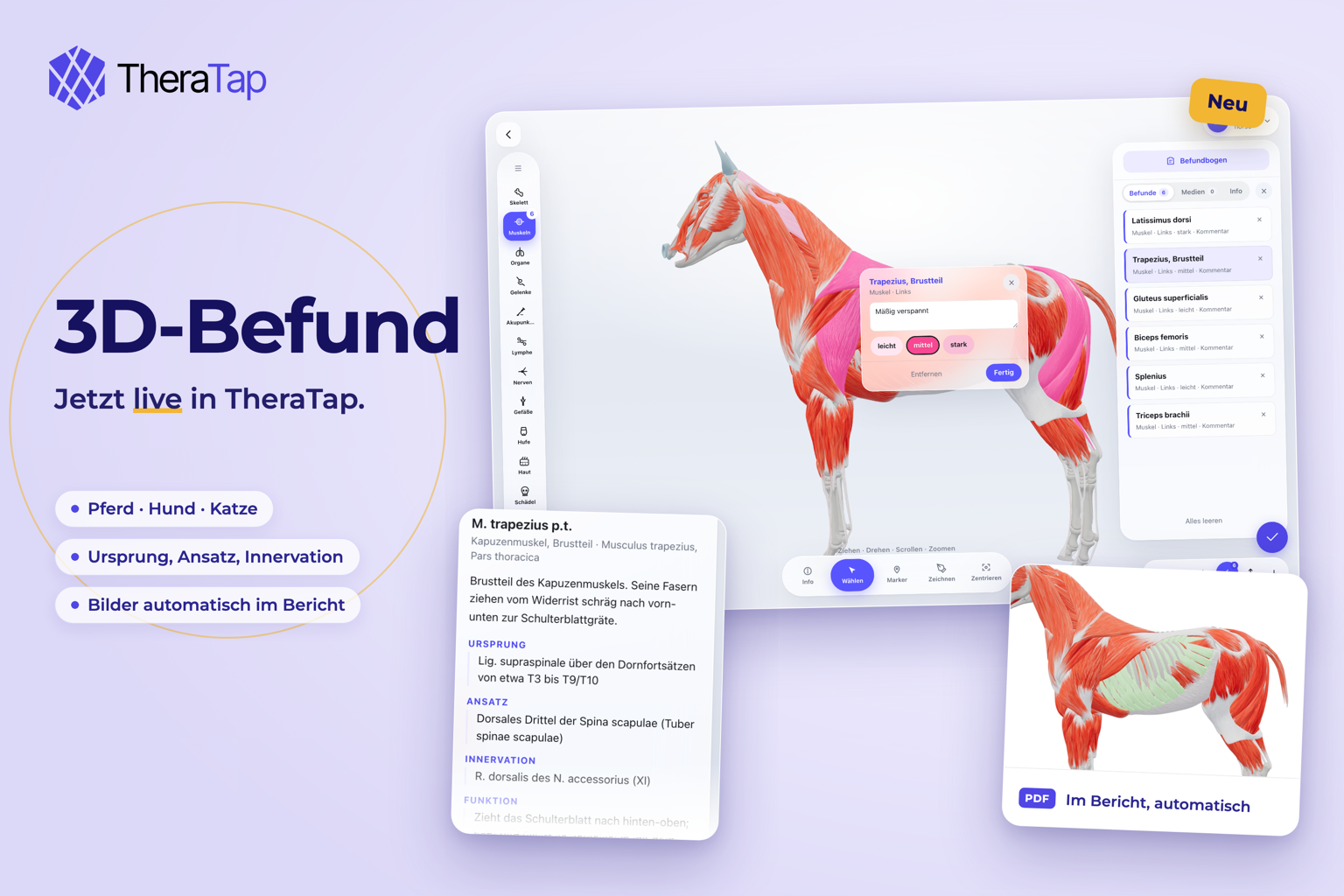
We announced it in September, now it is here: in TheraTap you record findings directly on the 3D model.
3D or 2D, your choice
The first time you click New assessment sheet, TheraTap asks once: 3D anatomy or 2D map? The answer becomes the default for your whole team and is shown on the button. The arrow next to it lets you pick the other type for a single assessment; change the default under Settings → Practice details. Existing assessments stay as they are – 2D assessments still open the familiar map.
Marking on the model
Horse, dog and cat come as complete 3D models: skeleton, muscles, organs, joints, lymph, nerves, vessels, hooves or paws, skin and acupuncture points. Rotate and zoom, show or hide layers, tap a part. As on the 2D map it is colored only once you enter a level, a rotation, a team option or a comment. Free markers and freehand strokes are available too.
Form, media, info
The familiar assessment form opens on top of the model. Drag photos and videos straight into the media tab. And the info tab shows origin, insertion, innervation, function and practice notes for every muscle, bone and organ – also without a finding, as a reference.
Report and reference content
For the PDF report, TheraTap creates images of the model automatically – one view per layer, framed on your markings. The reference content has been editorially checked; veterinary sign-off is still pending.
All details in the animal models guide.